Back Of Skull Anatomy - 3d male human skull bone. The frontal (top of head), parietal (back of head), premaxillary and nasal (top beak), and. Atlas of human skeletal anatomy. The skull begins to form prior to week 12 of embryogenesis. Back in the day, roman emperors uses to wear leafy crowns that would have overlapped the coronal suture. In order to be light, the skull is made up by flat and irregular bones, and has hollow spaces called the sinuses.
Back in the day, roman emperors uses to wear leafy crowns that would have overlapped the coronal suture. Atlas of human skeletal anatomy. The skull performs vital functions. The major sutures are the coronal suture, sagittal suture, lambdoid suture and squamosal sutures. Foramina inside the body of humans and other animals.
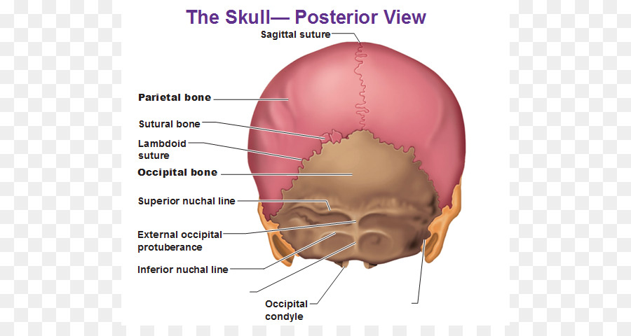
Human body Skull Anatomy External occipital protuberance Human back - Image Of Skull png ... from banner2.kisspng.com The skull bones can be classified into two groups: It serves aesthetic purposes since it provides the visible appearance of the face. Skull bones aren't fused together at birth. Excluding ear ossicles, it is made of 22 bones. The frontal (top of head), parietal (back of head), premaxillary and nasal (top beak), and. The skull encases and protects the brain as well as the special sense organs of vision, hearing, balance, taste and smell. So, the human skull consists of 23 bones. It supports and protects the face and the brain.
Learn more about the anatomy and function of the skull in humans and other vertebrates.
It is believed that trepanation was used to either relieve painful headaches, or to release demons from the skull. Learn about the anatomy of the skull bones and sutures as seen on ct images of the brain. Looking at it from the inside it can be subdivided into. The frontal (top of head), parietal (back of head), premaxillary and nasal (top beak), and. The occipital muscle is cupped like a saucer to accommodate the back part of the brain. Learn about skull base anatomy with free interactive flashcards. Skull bones aren't fused together at birth. The skull performs vital functions. The skull anatomy has many functions aside from protecting the brain. Overview, anterior skull base, middle skull base march 18, 2017. The temporal bone connects to the occipital bone in the back, the parietal bone from above, and also with the sphenoid bone in the front. Skull, skeletal framework of the head of vertebrates, composed of bones or cartilage, which form a unit that protects the brain and some sense organs. Human skull from the front.
The skull or known as the cranium in the medical world is a bone structure of the head. Continue scrolling to read more below. A cartilaginous mould begins to grow this is why raising your eyebrows can create the appearance that the back of the head is moving. A thorough description is beyond the. It is believed that trepanation was used to either relieve painful headaches, or to release demons from the skull.
Natural Birth In Kitsap: Optimal Fetal Positioning, Part 2 - The Fetal Head from 3.bp.blogspot.com A cartilaginous mould begins to grow this is why raising your eyebrows can create the appearance that the back of the head is moving. It supports and protects the face and the brain. The skull has a single occipital condyle.7 the skull consists of five major bones: The base of the skull (or skull base) forms the floor of the cranial cavity and separates the brain from the structures of the neck and face. Human skull from the front. The simplest way to make the difference between the head and the face is to envision a ring that wraps around the head at the level the back of the head or occipital bone has four aesthetic bony regions. 12 photos of the bone of back of skull. Looking at it from the inside it can be subdivided into.
The skull anatomy has many functions aside from protecting the brain.
The bbc is not responsible for the content of external websites. A thorough description is beyond the. The major sutures are the coronal suture, sagittal suture, lambdoid suture and squamosal sutures. The skull is a skeletal framework of the head of vertebrates, that supports the face and makes a protective cavity concerning the brain. Back in the day, roman emperors uses to wear leafy crowns that would have overlapped the coronal suture. Overview, anterior skull base, middle skull base march 18, 2017. Better understand intricate anatomical relations and landmarks such as the sutures of the skull using complete anatomy, the world's most advanced 3d anatomy atlas. It offers protection to the brain, eye balls, inner ears, and nasal passages. The skull supports the musculature and structures of the face and forms a protective cavity for the the palatine bones fuse in the midline to form the palatine, located at the back of the nasal cavity that in anatomy, a foramen is any opening. Foramina inside the body of humans and other animals. Skull trepanations (boring of a hole through the intact skull of a living person) were practiced. These joints fuse together in adulthood. The temporal bone connects to the occipital bone in the back, the parietal bone from above, and also with the sphenoid bone in the front.
Human skull from the front. The skull includes the upper jaw and the cranium. The skull bones can be classified into two groups: It offers protection to the brain, eye balls, inner ears, and nasal passages. Continue scrolling to read more below.
Back of skull labeled | Medical anatomy, Medical knowledge, Medical student study from i.pinimg.com The skull begins to form prior to week 12 of embryogenesis. The skull anatomy has many functions aside from protecting the brain. The skull or known as the cranium in the medical world is a bone structure of the head. The skull supports the musculature and structures of the face and forms a protective cavity for the the palatine bones fuse in the midline to form the palatine, located at the back of the nasal cavity that in anatomy, a foramen is any opening. Anatomy ▶ head and neck ▶ bones and cartilages ▶ skull. Continue scrolling to read more below. Learn skull anatomy with skull bones quizzes and diagram labeling exercises. Overview, anterior skull base, middle skull base march 18, 2017.
But it's not all bones!
12 photos of the bone of back of skull. It offers protection to the brain, eye balls, inner ears, and nasal passages. The frontal, parietal, temporal and occipital bones are joined at the cranial sutures. The skull is a bony structure that supports the face and forms a protective cavity for the brain. Overview, anterior skull base, middle skull base march 18, 2017. It is comprised of many bones, formed by intramembranous ossification, which are joined together by sutures (fibrous joints). The skull or known as the cranium in the medical world is a bone structure of the head. Continue scrolling to read more below. Some bones give shape to the face, others protect the brain. So, the human skull consists of 23 bones. The bbc is not responsible for the content of external websites. Foramina inside the body of humans and other animals. Skull reshaping is done on any of the structures that lie above the face.
Share :
Post a Comment
for "Back Of Skull Anatomy - 3d male human skull bone"


Post a Comment for "Back Of Skull Anatomy - 3d male human skull bone"